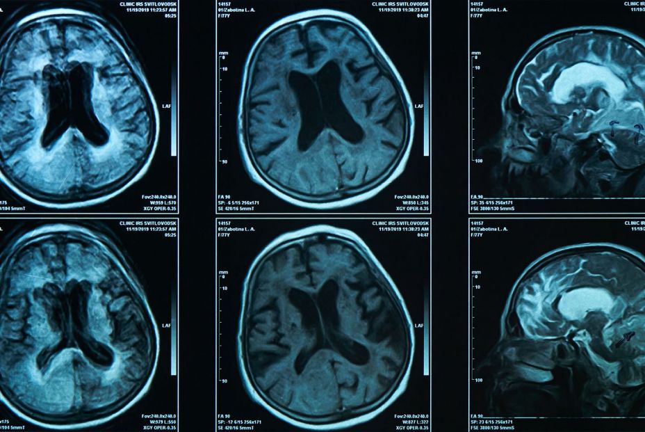
As the main cause of dementia worldwide, the disease of Alzheimer’s It represents a growing public health crisis. This disease is characterized by accumulation of abnormal proteins in the braincalled beta amiloid and tau, which manifest years before the appearance of clinical symptoms and can be detected by positron emission tomographies (PET).
Treatments aimed at these proteins have limited efficacy, indicating that other factors could contribute to cognitive impairment.
High iron levels in the brain are a factor that has been investigated in recent years. It is known that iron overload in the brain drives neurodegeneration by inducing oxidative stress (an imbalance between two types of molecules in the body: free and antioxidant radicals), which aggravates amyloid toxicity, alters the function of the Tau protein and promotes the death of nerve cells.
They detect iron levels in different regions of the brain
A special magnetic resonance technique that detects iron levels in different regions of the brain can predict the appearance of mild cognitive impairment and cognitive impairment in older adults without cognitive impairment, according to a study by the Johns Hopkins University (United States).
The results are published in ‘Radiology‘, a magazine of the North America Radiological Society (RSNA) And they point out that this could be a route to earlier interventions.
He brain iron It can be measured non -invasively through a special magnetic resonance technique called quantitative susceptibility mapping (QSM).
“QSM is an advanced magnetic resonance technique (RM) developed during the last decade to measure tissue magnetic susceptibility with high precision,” explains the main author of the study, Dr. Xu Li, associate professor of radiology at the Johns Hopkins University and associate researcher of the FM Kirby Research Center for functional images of the brain in the Kennedy Krieger Institute In Baltimore, Maryland.
“QSM can detect small differences in iron levels in different brain regions, which provides a reliable and non -invasive method to map and quantify iron in patients, something impossible with conventional RM methods.

Study in 110 patients
Dr. Li and his collaborators studied the magnetic resonance QSM in 158 participants without cognitive impairment, selected from the Biocard study of Johns Hopkins, a research project focused on Early stages of Alzheimer’s disease and related disorders. PET data of 110 of the participants was available.
The researchers obtained basal QSM magnetic resonance data from the participants and monitored them up to seven and a half years. They discovered that greater basal magnetic susceptibility in magnetic resonance in the enterorrinal cortex and putamen (two important brain regions for memory and other cognitive functions) was associated with a greater risk of mild cognitive impairment, a transition stage that precedes the dementia related to Alzheimer’s disease.
“Through the use of QSM, we find higher levels of brain iron in certain regions related to memory, which are linked to a greater risk of developing cognitive deterioration and faster cognitive deterioration,” says Dr. Li. “This risk is even higher when participants have higher levels of amyloid pathologies.”
Although the amyloid load and the tissue susceptibility in the entorrinal cortex and the putamen They associated independently with the progression to mild cognitive deteriorationThey seemed to have synergistic effects, accelerating global cognitive deterioration over time. If they are confirmed in broader studies with populations of more diverse patients, the findings point to a paper for magnetic resonance QSM in the evaluation of patients with risk of dementia.
“We can use this type of tool to help identify patients with the greatest risk of developing Alzheimer’s and, potentially, orienting early interventions as new treatments are available,” says Dr. Li. “In addition to serving as a biomarker, brain iron could become a future therapeutic target.”
In the future, researchers hope to better understand how cerebral iron contributes to Alzheimer’s disease, including its interaction with other pathologies related to this disease, such as amyloid and tau proteins. From the therapeutic point of view, clinical trials could evaluate therapies directed to iron. “At the same time, we hope that QSM technology is more standardized, faster and more accessible in clinical practice,” Li concludes.


